Average Reviews:

(More customer reviews)Very useful book, with the use of this book you can do EMG, botox and myobloc injection in hand, forarm and neck muscles very easely. Provides very good pictures of the muscles with their nerve supply and roots/segments. I start to use it in my clinic on patients that needed rare type of EMG testing muscles and it helps alot. This book shows and instruct you to find the muscle that you are looking for, the muscle primary fuction and movement. I highly recommend this book to whom wants to do EMG, botox or myoblock injection.
Click Here to see more reviews about: Atlas of Electromyography
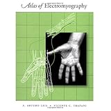
The Atlas of Electromyography is a visually alluring book which provides high quality anatomical illustrations of skeletal muscles that include nerve, plexus, and root supply; photographs of each muscle in healthy subjects to enable the practitioner to identify the optimum siteof EMG needle insertion; clinical features of the major conditions affecting peripheral nerves; and electrodiagnostic strategies for confirming suspected lesions of the peripheral nervous system. The atlas is divided into sections on the major peripheral nerves. Each nerve is illustrated and its anatomy reviewed in the text. The authors provide a detailed outline of the clinical conditions and entrapment syndromes that affect the nerve, including a list of etiologies, clinical features, and electrodiagnostic strategies used for each symdrome. Each muscle supplied by the peripheral nerve is shown as an anatomical illustration with a corresponding human photograph. The text provides information about the muscle origin, tendon insertion, voluntary activation maneuver, and site of optimum needle insertion. The needle insertion point is identified in both the anatomical illustration and the corresponding photographs. This assures that pertinent bone, muscular, and soft tissue landmarks can be used to guide the electromyographer to a specific point on the skin. Potential pitfalls associated with the needle insertion are added, usually noting adjacent muscles or structures that may be mistakenly entered. Clinical correlates pertinent to the muscle being examined are also provided. The tlas of Electromyography serves as an anatomical guide for practitioners of electromyography and neurologists, as well as residents i neurology, physical medicine, and rehabilitation.
Click here for more information about Atlas of Electromyography

No comments:
Post a Comment